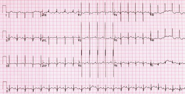
Below is an ECG taken of a 0ne week old infant, who has some shortness of breath whilst feeding according to the parents. What does it show? Is it normal?
ECG’s can already present a significant challenge, however there is a fear associated with the paeds ECG. There should not be, as the same principles that apply to the adult ECG and the use of the ‘ECG in 20 Seconds Approach’, also apply to children.
There are only 3 things to remember, in fact there is only one new thing to remember in a paediatric ECG i.e.., In young children, the right ventricle is dominant. As they grow, the left ventricle becomes dominant. That’s it. Everything else follows.
Here is the approach as per the ‘ECG in 20 Seconds’
Rate: just count the number of complexes and multiply by 6 ( an ECG takes 10 seconds to print).
What is an appropriate rate for children? I use this very rough rule of thumb and usually also look it up:
- < 2yo max HR is 160
- 2-7yo max 140
- 7-15 yo max 130
Rhythm: make sure that it is sinus. For sinus rhythm, there must be a P wave before each QRS and the P wave must be upright in II and inverted in aVR. If not, then it is not sinus rhythm and the pacemaker is in the low right/left atrium
QRS: is it too tall or too small, ie is there hypertrophy? Remember that in the child the right ventricle is bigger than the left. To judge hypertrophy look at V6. If the R wave intersects the baseline of the V5 tracing above, then there is LVH
In terms of Right ventricular hypertrophy, in adults it is usually hidden by the left ventricle, however in children the clues are in V1. There is RVH if there is an rsR pattern in V1, or a pure R wave in V1 after 6 months.
It is also helpful to look at the R wave to S wave ratio. Remember that the young heart is right heart dominant, so you would expect that the R wave is more predominant than the S wave in the right leads, however if the opposite is true, then there is left ventricular hypertrophy.
The QRS is normally < 1 small box i.e.., 0.04sec. If longer than 2-3 small squares it is a bundle branch block.
The QRS is normally +ve in aVF
- If it is negative in aVF, there may be cardiac malformations, such as AV septal defect.
- If it is biphasic in aVF, it may be normal, but needs cardiology discussion.
Look for the abnormal QRS morphology i.e.., the delta wave of WPW
ST-T: The T wave is upright for the first week of life, then is inverted until adolescence. This is normal.
Intervals:
PR- In anyone, an interval > 0.2 sec is abnormal
- In infants and young children an interval of > 0.16 (4 small squares) is abnormal.
QT- >0.45sec is abnormal in anyone
- In < 6 months <0.49sec
- In > 6 months <0.44
Lets go back to the ECG above:
Rate: 23 complexes x 6 = 138
Rhythm: There are P waves before each QRS and they are upright in II and inverted in aVR. This normal sinus rhythm.
QRS: Not too narrow or too wide i.e. about 1 small square. No abnormal morphology. No LVH.
ST-T: the one obvious characteristic is inverted T waves in the anterior leads. This is normal in children between about 1 week and adolescence.
Intervals: PR is normal, QT is normal.
This is normal sinus rhythm.
Lets look at some differences:
Rate and rhythm you know; how about:
QRS
This ECG is of an infant with Downs Syndrome
Look at the QRS in V1- there is an rsR pattern indicating right ventricular hypertrophy. This patient had primary pulmonary hypertension.
How about this one of a 10 year old ( Am J Emerg Med (2008) 26, 221-228):
Note that the QRS are the right width, however there is a pure R wave in V1 indicating RVH. If we look at aVF the QRS is biphasic. This may be abnormal, certainly abnormal if the QRS is -ve in this lead. The T waves are inverted as we would expect in a 10 yo. This is right ventricular hypertrophy secondary to primary pulmonary hypertension.
I hope that helps.
If you want to get better at the ECG come to one of our ECG workshops. In 3 hours you’ll be happy about ECG’s.


How to Read The Paediatric ECG – Resus
[url=http://www.g63t8q288n6a1j25mehx2uo645zn2ni0s.org/]ujpdjszyik[/url]
ajpdjszyik
jpdjszyik http://www.g63t8q288n6a1j25mehx2uo645zn2ni0s.org/
Car Brake Caliper
iphonexrブランドコピー
Large Telehandler
Gucciグッチ靴スーパーコピー
Aluminium Door Profile
Gucciグッチ財布スーパーコピー
Hermesエルメスバッグスーパーコピー
best male vibrator
44b Bras Plus Size
LouisVuittonルイヴィトンイヤリングスーパーコピー
SaintLaurentサンローラン財布スーパーコピー
Tea Bag Paper
Engine Water Pump
Chanelシャネルベルトスーパーコピー
Acme Transformer
Gucciグッチバッグスーパーコピー
Inner Tie Rod End
スーパーコピーブランド
Fendiフェンディ帽子スーパーコピー
Heavy Metal- Freepvc Particle
Cng Cylinder Types
LouisVuittonルイヴィトンマフラー販売店
Cargo Elevator
Hermesエルメスイヤリング販売店
Oil Free Makeup Remover Wipes
ブランドDiorディオールサングラスコピー代引き
Land Rover Auto Brake Pad
日本国内最高級スーパーコピーブランド激安販売
Kubu Wicker Baskets
ブランドコピー代引き
スーパーコピーバッグ
China Tote Bag and Totes price
Stainless Steel Woven Wire Mesh
ブランドバッグコピー
Vibrating Screen
ブランドGucciグッチサングラスコピーN級品
Led Display Module
Celineセリーヌコピー激安
portable site toilet for sale
ブランドバッグコピー
Goyardゴヤールコピー激安
Wall Plastering Machine Rendering
China 40k Fat Cavitation
RogerVivierロジェヴィヴィエ靴販売店
ブランドコピー専門店
Endotracheal Intubation Ppt
ブランド時計コピー
2000l Brew House Electric Heating
QSFP+ 2km Supplier
ブランドFendiフェンディ帽子コピー代引き
Home Diesel Generator
Balenciagaバレンシアガバッグコピー
ブランド財布コピー
China Zhuji Valve Factory
ブランドコピーFendiフェンディN級品
Hand Sanitizer Bottle
SaintLaurentサンローラン財布販売店
others
スーパーコピーブランド
High Quality Digital Portable Slit Lamp Microscope