A 23 year old male presents with 6 hours of central sharp chest pain, left arm heaviness and diaphoresis. He has no past medical history or relevant family history. He doesn’t smoke and is a social drinker. He denies any elicit substances. His vitals are: HR as per ECG, BP 128/67, Sats 97% on RA.
His ECG is shown below.
Describe the ECG and answer the following question:
If this patient presented to a rural emergency department and you did not have a cardiac cath lab, would you thrombolyse?
Describe the ECG as if were sitting the Fellowship Exam
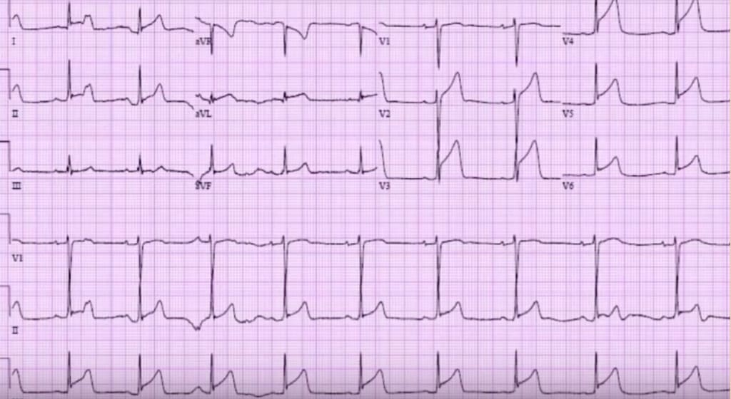
RATE: 6x 9 complexes = 54
P WAVES: Upright in II and inverted in aVR, so sinus. No extra p’s
QRS’s: Not too tall/small, wide/narrow or abnormal morphology.
AXIS: Normal
ST-T SEGMENTS: There is ST elevation in V2-V6, I, II, aVL, however, there are no reciprocal changes. There is a QRT sign where the T wave shoots straight up, that may indicate ischaemia. The T wave is as the R wave in V4 again indicating ischaemia.
T-P SEGMENT: In a couple of the leads there is a small downsloping of the T-P segment which may be indicative of a Spodick’s Sign
PR/QT INTERVALS: All normal, although there are some leads with PR depression
PACING SPIKES: No pacing to be seen
Is this an ischaemic ECG and would you Thrombolyse
There is diffuse ST elevation. If you look closely there is some PR elevation in aVR(may indicate pericarditis)but lets ignore because evidence is poor. There may also be some PR depression in II.What are the things that strongly favour STEMI?
- ST depression in any lead except aVR or V1- NONE here
- STE III>II- NOT here
- Horizontal or convex ST elevation – there is some straightening T waves in aVL
- QR-T sign- as the R wave is descending, it then goes up wards, without forming any ST segment. PRESENT
- The T wave “towers” over the R wave in V4- very strong sign
What Happened?
This ECG favours a STEMI. Yes even in a 23 year old.Following Cardiac Consult he would more than likely receive thrombolysis.
As luck would have it for this young man, the retrieval service was on the ground in our hospital so he got choppered to a hospital with a cath lab.
THE RESULT
- 99% prox LAD stenosis
- 100% distal LAD stenosis


ECG of the week 22 Februrary 2017 – Resus
[url=http://www.gg263ho63699jaygphw497u15ge3o28zs.org/]ugnekdyyqqk[/url]
gnekdyyqqk http://www.gg263ho63699jaygphw497u15ge3o28zs.org/
agnekdyyqqk