How good are you at reading elbow X-rays? Here are some tools and rules that will help you pick up those elbow fractures.
Fractures of the elbow usually fall into the following groups:
- CHILDREN: Supracondylar fractures
- ADULTS: Radial Head Fractures
- ELDERLY: Multiple Fractures
Fractures are a CLINICAL Diagnosis. The x-ray merely confirms that you have found it. A fracture can occur with a normal X-ray.
HINT 1: If the patient presents with clinical signs of a fracture and the x-ray is normal, treat them like there is a fracture.
What are the Clinical signs?
- Swelling
- Deformity
- Point tenderness
- Examination of range of motion.
- Perform the elbow extension test.
What are the X-ray Signs?
The Fat Pad Sign
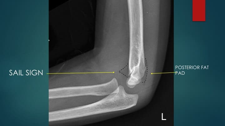
If you can see a fat pad, it simply indicates a joint effusion. You might normally see the anterior fat pad. However when it is so displaced by an effusion as to result in the ‘sail sign’ or when you see the posterior fat pad, it is a good marker for a fracture.
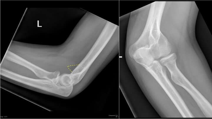
CASE
This is a 32 yo woman who fell off her bicycle and landed on her elbow, presents with pain in the elbow and limited movement. The X-rays are shown below:
Its obvious that there is a sail sign, but there is no obvious fracture. What now?

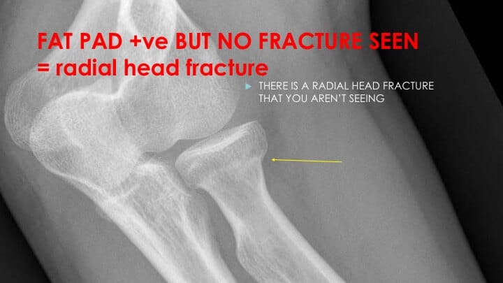
HINT 2: If there is a positive fat pad sign but no fracture seen in an adult, think of a radial head fracture, it will be there.
Anterior Humeral Line
The anterior humeral line, assists us in identifying normal elbow alignment but also in diagnosing a supracondylar fracture in children.
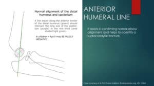 A line is drawn along the anterior surface of the humerus. In the normally aligned joint, it should pass through the middle third of the capitellum.
If it doesn’t pass through this point then there is abnormal alignment of the elbow joint. In a supracondylar fracture this can occur due due posterior displacement of the distal humerus.
Beware in children less than 4 years of age there may be false negatives.
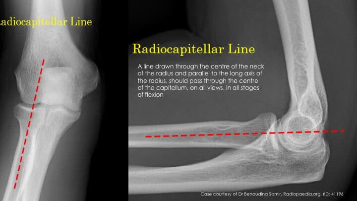
Radiocapitellar line
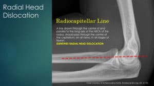 It is a helpful line as it identifies radial head dislocation. This is a line drawn through the centre of the radius, running parallel to the neck of the radius. It should pass through the centre of the capitellum. One of the common mistakes, is to draw the line parallel to the shaft of the radius and not to the neck of the radius.
It should pass through the capitellum in all views
If we apply this to the previous case:
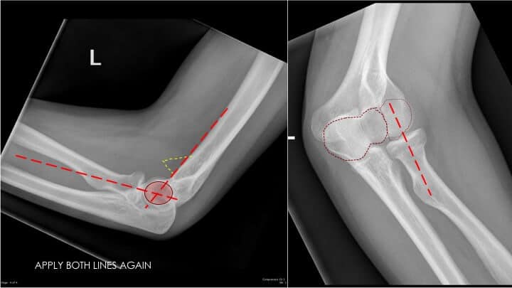
The line passes parallel to the neck of the radius. Both the anterior humeral line and the radiocapitellar line pass through the centre of the capitellum. It tells us there is normal alignment of the joint, but doesn’t tell us about a fracture. We know the patient had a radial head fracture.
My 4 Rules for reviewing the elbow X-ray to not miss any.
- If the patient presents with clinical signs of a fracture and the x-ray is normal, treat them like there is a fracture.
- Deformity and swelling
- Point tenderness
- Range of movement
- Elbow extension Test
- If there is a positive fat pad sign but no fracture seen in an adult, think of a radial head fracture.
- Draw an Anterior Humeral Line, to assess joint alignment.
- Draw a Radiocapitellar Line, to exclude a radial head dislocation.
Peter Kas